Introduction:
The medical ailment known as adhesive capsulitis, or frozen shoulder, is characterized by discomfort and stiffness in the shoulder joint. If not properly controlled, it can seriously impair a person’s capacity to carry out everyday tasks and result in persistent impairment. This article examines the pathophysiology, manifestations, etiology, evaluation, and all-encompassing care of frozen shoulder.

Pathophysiology:
The thickening and inflammation of the shoulder capsule, and the connective tissue around the shoulder joint, are hallmarks of a frozen shoulder. It is thought to entail a combination of fibrotic and inflammatory processes, while the precise mechanism is yet unclear. Scar tissue is created as a result of inflammation, and this limits the range of motion in the joint. This can be triggered by systemic disorders, including diabetes, extended immobilization, or trauma.
Stages of Frozen Shoulder:
Frozen shoulder generally progresses through three stages:
- Freezing Stage (Painful Stage)
- Duration: 6 weeks to 9 months.
- Characteristics: Gradual onset of pain, worsening over time, often severe at night, and limiting shoulder movement.
- Frozen Stage (Stiffening Stage)
- Duration: 4 to 12 months.
- Characteristics: Pain may lessen, but stiffness increases significantly, making daily activities difficult.
- Thawing Stage (Recovery Stage)
- Duration: 6 months to 2 years.
- Characteristics: Gradual improvement in shoulder range of motion, with decreasing pain and a slow return to normal function.
Causes:
The precise cause of a frozen shoulder is often unclear, but several factors can contribute to its development:
- Idiopathic: Occurs without a known cause.
- Trauma or Surgery: Injuries or surgical procedures involving the shoulder can lead to immobilization, increasing the risk of adhesive capsulitis.
- Systemic Conditions: Frozen shoulder has been associated with an increased risk of diabetes, thyroid diseases, cardiovascular disease, and Parkinson’s disease.
- Prolonged immobilization: Shoulder injuries such as fractures that prevent movement for extended periods of time might result in a frozen shoulder.
Assessment:
1. Medical History
A comprehensive medical history should include:
- Onset of Symptoms: Determine the onset and whether symptoms began gradually or suddenly.
- Duration and Progression: Note the duration of symptoms and any changes over time.
- Pain Characteristics: Assess the location, intensity, quality, and pattern of pain.
- Previous Shoulder Injuries or Surgeries: Document any history of shoulder trauma or surgeries.
- Medical Conditions: Check for systemic conditions associated with a frozen shoulder.
- Lifestyle Factors: Consider occupational and recreational activities that may contribute to shoulder problems.
2. Physical Examination:
The physical examination should focus on evaluating the shoulder’s range of motion, strength, and overall function:
- Inspection:
- Observe for visible deformities, swelling, or muscle atrophy.
- Compare the affected shoulder with the unaffected shoulder.
- Palpation:
- Identify areas of tenderness, warmth, or swelling.
- Assess the acromioclavicular joint, biceps tendon, and rotator cuff insertions.
- Range of Motion (ROM) Assessment:
- Active ROM: Ask the patient to perform shoulder movements (flexion, extension, abduction, adduction, internal rotation, external rotation) and document limitations or pain.
- Passive ROM: Move the patient’s shoulder through the same motions while they remain relaxed, noting any restrictions.
- Strength Testing:
- Evaluate the strength of the deltoid, rotator cuff muscles, and scapular stabilizers.
- Compare strength bilaterally.
- Special Tests:
- Apley Scratch Test: Assess shoulder mobility by having the patient reach behind their back.
- Neer and Hawkins-Kennedy Tests: Differentiate between frozen shoulder and rotator cuff pathology.
- Lift-Off Test: Evaluate subscapularis muscle function by asking the patient to place their hand on the lower back and lift it off against resistance.
3. Imaging Studies: Imaging studies confirm the diagnosis and rule out other conditions:

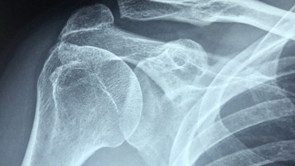
- X-rays: Standard shoulder X-rays (anteroposterior, lateral, and axillary views) rule out fractures, dislocations, and significant arthritic changes.
- Magnetic Resonance Imaging (MRI): enables the identification of abnormalities such as the thickness of the joint capsule, inflammation, and other details through comprehensive pictures of the soft tissues.
- Ultrasound: Assesses the rotator cuff tendons and detects associated bursitis or tendonitis, joint effusion, and synovial hypertrophy.
4. Differential Diagnosis:
Differentiate frozen shoulder from other conditions with similar symptoms:
- Rotator Cuff Tears: Present with pain and weakness in specific movements.
- Subacromial Bursitis: Characterized by pain during shoulder movements, especially overhead activities.
- Osteoarthritis: Involves pain, crepitus, and gradual loss of joint space visible on X-rays.
- Cervical Radiculopathy: Presents with referred shoulder pain and neurological symptoms like numbness or tingling in the arm.
Comprehensive Management of Frozen Shoulder:
Non-Surgical Management:

- Patient Education and Activity Modification:
- Education: Provide details about the illness, how it usually resolves, and how crucial it is to follow the prescribed course of action.
- Activity Modification: Advise avoiding activities that exacerbate pain, while encouraging gentle shoulder movements.
- Pain Management:
- Medications: Prescribe NSAIDs (e.g., ibuprofen, naproxen) for pain and inflammation, and consider analgesics like acetaminophen.
- Corticosteroid Injections: Inject intra-articular corticosteroids to relieve pain and decrease inflammation, particularly in the early stages.
- Physical Therapy:
- Range of Motion Exercises: Begin with passive stretching, progressing to active-assisted and active stretching as pain decreases.
- Strengthening Exercises: Introduce isometric exercises, progressing to isotonic exercises to strengthen the rotator cuff and scapular stabilizers.
- Manual Therapy: Myofascial release methods, soft tissue massage, and joint mobilizations should all be included.
- Heat and Cold Therapy:
- Heat Therapy: Apply moist heat packs before exercises to relax muscles and increase blood flow.
- Cold Therapy: Use ice packs after exercises to reduce inflammation and pain.
- Hydrodilatation:
- A procedure involving the injection of saline mixed with corticosteroid and anesthetic into the joint capsule to stretch it and improve mobility.
Spencer Techniques:
Spencer techniques are a series of osteopathic manipulative techniques used to improve mobility and reduce pain in the shoulder joint. They include:
- Traction with the Arm Extended: Applying traction to the shoulder joint with the arm extended.
- Traction with the Elbow Flexed: Applying traction with the elbow flexed.
- Circumduction with Compression and Traction: Moving the shoulder in circular motions with applied compression or traction.
- Circumduction with Traction (Elbow Flexed): Circular motions with traction and the elbow flexed.
- Abduction with Traction: Moving the shoulder into abduction while applying traction.
- Internal Rotation: Applying internal rotation to the shoulder joint.
- Traction with Superior-Inferior Glide: Applying superior-inferior glide while maintaining traction.
- Abduction with Internal Rotation: Combining abduction and internal rotation movements.
Joint Mobilization Techniques:
Different glides can help improve specific movements and reduce stiffness in the shoulder joint. These include:
- Anterior Glide: For shoulder extension and external rotation.
- Posterior Glide: For shoulder flexion and internal rotation.
- Inferior Glide: For shoulder abduction.
- Lateral Distraction: To increase overall joint mobility.
Surgical Management:
- Manipulation Under Anesthesia (MUA):
- Procedure: The shoulder joint is manipulated under general anesthesia to break up adhesions and improve mobility.
- Indications: Consider MUA for patients not responding to non-surgical treatments after 6 months or with severe limitations.
- Arthroscopic Capsular Release:
- Procedure: Minimally invasive surgery to cut tight portions of the joint capsule, releasing adhesions and improving range of motion.
- Indications: Recommended for patients with refractory frozen shoulder not responding to extensive non-surgical management and MUA.
Alternative Therapies:
- Acupuncture: May reduce pain and improve function by inserting fine needles into specific body points.
- Transcutaneous Electrical Nerve Stimulation (TENS): Uses electrical currents to provide pain relief by stimulating nerves.
- Hydrotherapy: Exercises in warm water can reduce pain and improve mobility.
Post-Surgical Rehabilitation:
- Early Mobilization: Initiate gentle range of motion exercises immediately after surgery.
- Gradual Progression: Increase exercise intensity and range as tolerated.
- Regular Follow-Up: Schedule follow-up visits to monitor progress and adjust the rehabilitation program.
Psychological Support:
- Counseling: Provide psychological support for coping with chronic pain and functional limitations.
- Stress Management: Encourage stress-reducing activities like yoga, meditation, and relaxation techniques.
Monitoring and Evaluation
- Regular Assessments: Monitor pain levels, range of motion, strength, and overall function through clinical assessments and patient-reported outcome measures.
- Outcome Measures: Utilize standardized outcome measures to evaluate the effectiveness of treatment, such as the Constant-Murley Score and the Shoulder Pain and Disability Index (SPADI).
Lifestyle Modifications:
- Diet and Nutrition: Encourage a balanced diet rich in anti-inflammatory foods and advise maintaining a healthy weight.
- Exercise and Fitness: Promote regular, low-impact exercises and encourage gentle stretching and strengthening exercises.
Conclusion:
A thorough, customized strategy is needed for the effective treatment of frozen shoulder. Enhanced results can be achieved by combining physical therapy, pain management, education for patients, and, if required, surgery. Restoring shoulder function and improving the quality of life for people with frozen shoulder requires early intervention, continuous monitoring, and a multidisciplinary approach. Optimal recovery depends on the treatment plan being customized to the patient’s unique demands and the stage of the ailment.
FAQs:
What is a frozen shoulder?
Adhesive capsulitis, another name for frozen shoulder, is a disorder that causes discomfort and stiffness in the shoulder joint. It happens when the capsule that surrounds the shoulder joint gets thicker and tighter, limiting movement.
What are the causes of frozen shoulder?
Although the precise cause of frozen shoulder is uncertain, the following are common associations:
Accident or Surgery: Shoulder trauma or surgery can cause a restricted range of motion, which can exacerbate a frozen shoulder.
Medical Conditions: A number of illnesses can raise the risk, including diabetes, thyroid issues, heart disease, and Parkinson’s disease.
Immobility: Extended immobility following surgery, a stroke, or a broken arm can result in a frozen shoulder.
How much time does recovering from frozen shoulder take?
The illness usually goes away with time, although a complete recovery may take several months to two or three years.
Treatment and physical therapy can hasten the healing process.
Which exercises are beneficial for treating frozen shoulder?
The following are some exercises that can aid with shoulder mobility:
Pendular exercises: Gently swing your arm in a circular motion.
Towel Stretch: Gently stretch while using both hands to hold a towel behind the back.
Finger Walk: To release tension in the shoulder, walk your fingertips up a wall.
Cross-Body Reach: Pull the afflicted arm gently across the body with the unaffected arm.


3 thoughts on “Frozen Shoulder: A Comprehensive Guide.”